Essential Screening Methods for Early Cancer Detection
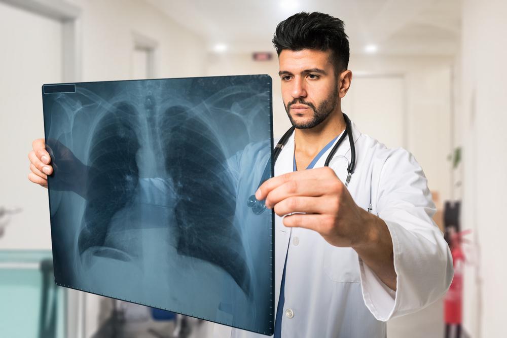
Early cancer detection is essential for successful treatment. Key diagnostic methods include imaging scans like CT, MRI, X-rays, nuclear medicine scans, ultrasounds, and minimally invasive procedures such as endoscopy and biopsy. These tools help identify tumors, determine their stage, and guide treatment decisions, ultimately saving lives and reducing healthcare costs.
Sponsored
Detecting cancer early is crucial for effective treatment and improved survival rates. Typically, these tests are performed when symptoms emerge, helping to identify cancer at an initial stage, which can reduce patient discomfort and healthcare costs. The American Cancer Society provides comprehensive screening guidelines that healthcare providers and patients should follow. Below are some key diagnostic procedures recommended for early cancer detection.
Imaging Techniques
Imaging procedures allow physicians to visualize internal body structures, aiding in the assessment of tumor size, location, and spread. Techniques include ultrasound, X-rays, CT scans, and MRIs, which provide detailed images to evaluate the severity and progression of cancer.
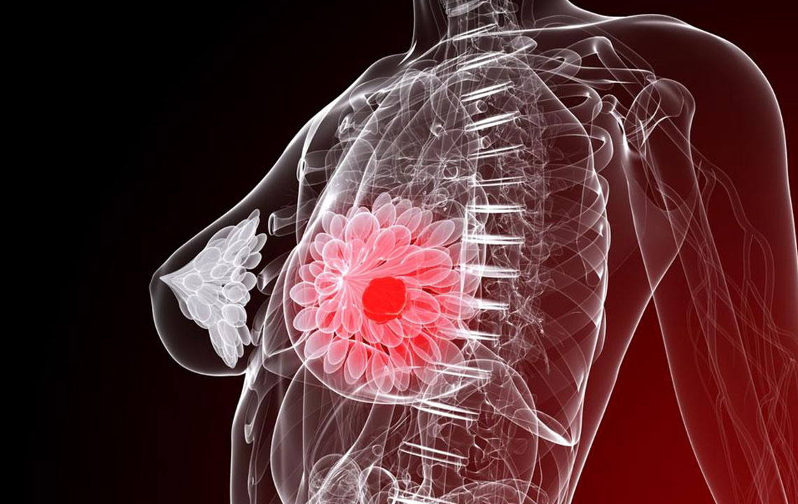
Imaging tests are vital for distinguishing benign from malignant tumors, guiding treatment planning, and staging cancer accurately. They also monitor tumor response after therapy.
CT Scans
Computed tomography scans, also called CAT scans, are non-invasive and painless. They generate cross-sectional images of the body's bones, organs, and tissues using radiation from multiple angles. This helps in precisely locating and measuring tumors and is used in treatments like radiofrequency ablation (RFA).
MRI Scans
Magnetic resonance imaging employs magnetic fields to produce detailed images of soft tissues and internal organs. Contrast dyes like gadolinium enhance clarity and help determine the origin and spread of cancer. Patients lie inside a large magnet, and radiofrequency waves generate the images.
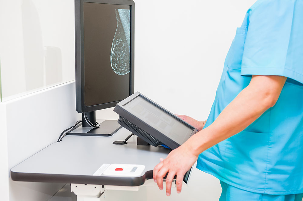
X-ray & Contrast Studies
X-rays create shadow images of bones and tissues, useful for early detection of certain cancers like breast cancer via mammograms. Contrast studies, involving dyes such as barium, improve visualization of internal structures, which is beneficial in diagnosing urinary or gastrointestinal cancers.
Nuclear Medicine Imaging
Nuclear scans involve radioactive substances called radionuclides to produce images based on cellular chemistry. These scans reveal areas with abnormal tissue activity, helping locate cancerous spots. However, they are less effective for detecting small tumors and are used alongside other diagnostic tools.
Ultrasound Examination
Ultrasound uses high-frequency sound waves to visualize organs, assess blood flow, and distinguish between cysts and solid tumors. It is radiation-free, quick, and effective for real-time imaging, especially for soft tissue evaluation.
Endoscopic Procedures
Endoscopy involves inserting a flexible tube with a light and camera into the body to examine internal organs directly. Techniques like colonoscopy are used to detect colorectal cancer, while laser and photodynamic therapies can treat tumors during the procedure.
Biopsy
A biopsy involves removing tissue samples for microscopic examination, often performed after imaging indicates abnormal growth. Fine-needle aspiration biopsy is a common minimally invasive method where a thin needle extracts cells for analysis.
Cellular and Fluid Analysis
Cytology examines individual cells obtained by scraping, brushing, or fluid collection from body sites like urine or respiratory secretions. It aids in diagnosing cancer, infections, or abnormalities such as cervical lesions via Pap smears.