Understanding How Rheumatoid Arthritis Is Diagnosed
This article provides a comprehensive overview of how rheumatoid arthritis is diagnosed. It covers symptoms, blood tests, and imaging techniques used by healthcare professionals to confirm the condition. Understanding these diagnostic methods is crucial for early detection and effective management of RA, preventing joint damage and deformity.
Sponsored
Rheumatoid arthritis (RA) is an autoimmune condition triggering joint inflammation. Here's what you should know about its diagnosis.
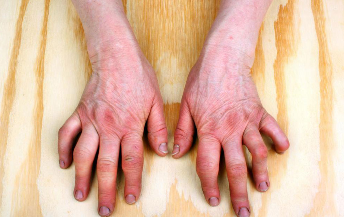
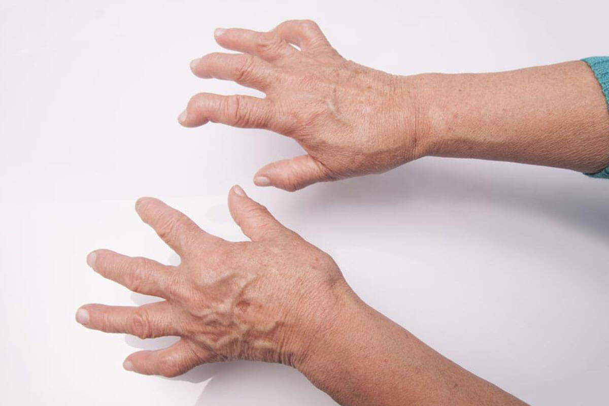
RA primarily affects small joints of the hands and feet, impacting roughly 1.5 million Americans. If untreated, it can damage cartilage and lead to joint deformities. It is more prevalent in women and typically occurs in those over 40. Beyond joints, RA can affect organs like the eyes, skin, lungs, and blood vessels.
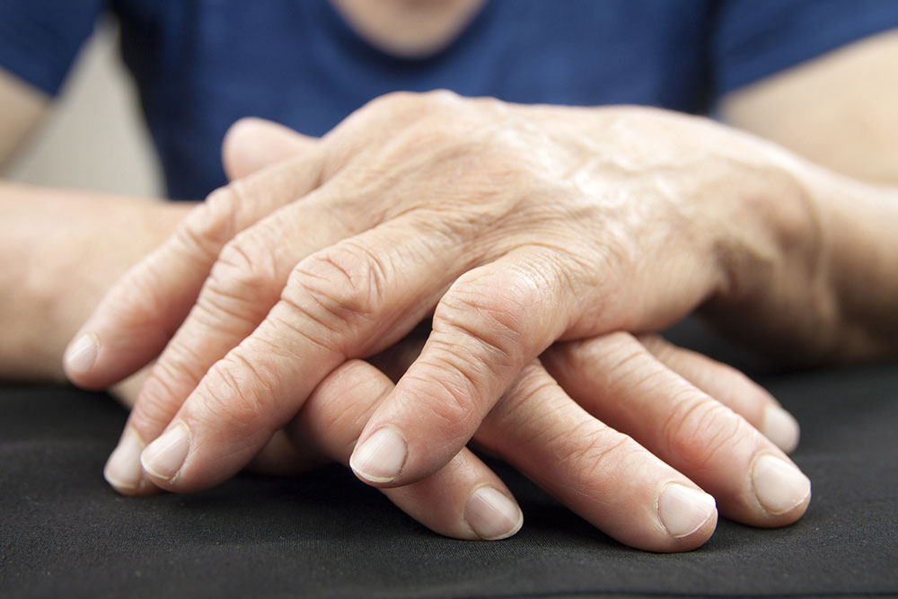
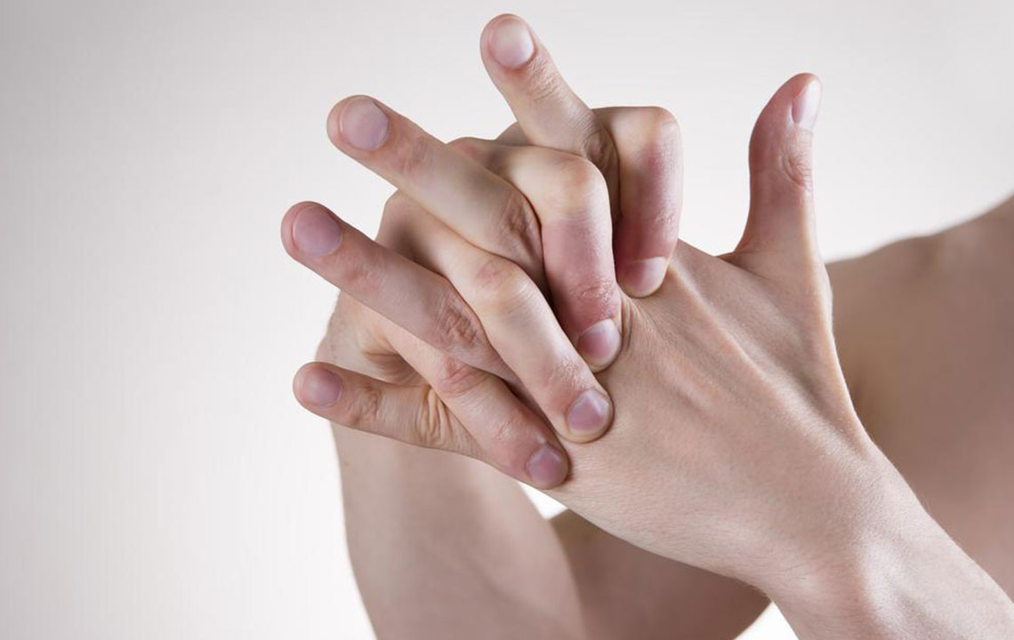
Common symptoms include fatigue, fever, morning stiffness, swollen joints, and weight loss. Symptoms often appear symmetrically, affecting both sides of the body. The severity varies, with periods of flare-ups and remission. Over time, RA can cause joint misalignment and deformity.
No definitive cause of RA is identified yet. Treatment focuses on alleviating pain and reducing joint stiffness after diagnosis. Diagnosing RA can be challenging, as other conditions cause similar symptoms. There’s no single test for RA, so doctors perform physical exams to check for signs like swelling, redness, and warmth in joints, along with muscle strength and reflex assessments. Confirmatory tests include blood analyses and imaging scans such as:
Blood Tests
No single blood test confirms RA definitively. Tests can indicate inflammation or autoimmune activity, including:
CRP (C-reactive protein) Test
CRP is produced by the liver in response to inflammation. Elevated levels suggest inflammation presence but don’t confirm RA specifically.
Rheumatoid Factor (RF) Test
This test detects RF antibodies, which are present in many RA cases but can also be positive in other conditions. Its results are suggestive but not conclusive.
Complete Blood Count (CBC)
CBC assesses anemia, common in RA patients. However, anemia alone isn't diagnostic for RA.
Joint Imaging Procedures
Imaging helps assess joint damage and differentiate RA from other types of arthritis. Common methods include:
Ultrasound – Uses sound waves to visualize joint inflammation.
X-Rays – Employ radiation to examine joint structure.
MRI – Uses magnetic fields to produce detailed images of joints.
These are essential tools for diagnosing RA and monitoring its progression.