Understanding Lung Damage in COPD: Key Pathological Features
This article explains the pathological mechanisms of COPD, including airway inflammation, mucus overproduction, and alveolar destruction. It highlights how these changes impair oxygen and CO2 exchange, worsening respiratory function in affected individuals.
Sponsored
Lung Damage in Chronic Obstructive Pulmonary Disease (COPD)
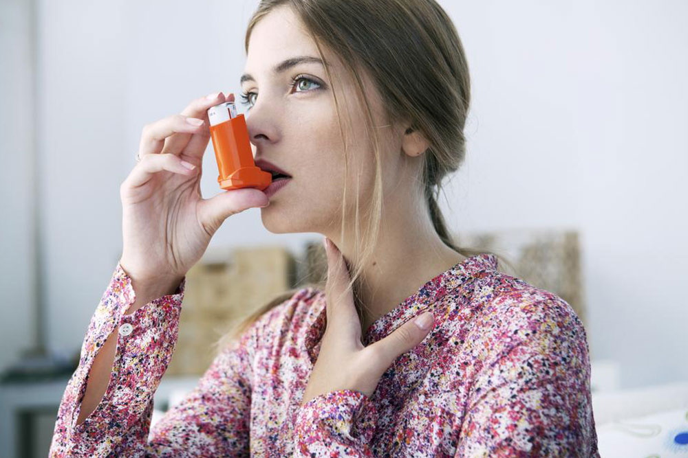
In healthy lungs, oxygen-rich air flows through the respiratory tract into tiny air sacs called alveoli, where oxygen is absorbed into the blood, and carbon dioxide is expelled. Air enters via the nose or mouth, travels through the trachea and bronchi, reaching the alveoli. In COPD, airflow is obstructed due to damaged alveoli and narrowed airways, impairing oxygen intake and CO2 removal. These changes include inflammation, mucus build-up, and alveolar wall destruction, which compromise effective gas exchange and respiratory function.
Chronic Bronchitis: characterized by inflamed, narrowed airways with excess mucus and swelling. The bronchial walls spasm, producing more mucus that clogs airways, reducing airflow. The inflammation damages the lining, worsening airway constriction.
Emphysema: involves destruction of alveolar walls, causing the alveoli to lose elasticity. This leads to impaired oxygen and carbon dioxide exchange because the alveoli cannot properly inflate or deflate, trapping air and decreasing efficiency.
Oxygen is essential for cell survival, and CO2 is a metabolic waste. Alveolar dysfunction: in emphysema, alveoli remain abnormally inflated due to blocked air exits. Normal alveolar function: inhaled oxygen fills the alveoli, and exhaled CO2 leaves. In COPD, impaired airflow causes residual CO2 in alveoli, mixing with fresh air, lowering oxygen intake. Alveolar destruction: occurs because enzymes like elastase damage elastin in alveoli, weakening their structure, similar to an old, worn-out balloon.