Essential Facts About Knee MRI Imaging
Learn everything about knee MRI scans, including their purpose, how they work, conditions they diagnose, and safety considerations. This non-invasive imaging technique is crucial for detecting knee injuries and abnormalities with high precision, helping healthcare providers plan effective treatment strategies while understanding potential risks involved.
Sponsored

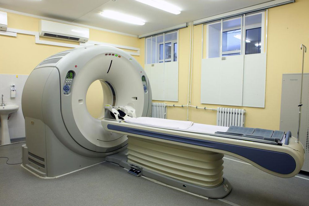
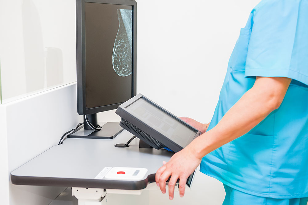
An MRI (Magnetic Resonance Imaging) scan uses powerful magnets and radio waves to generate detailed images internally, without surgery. This imaging technique is applicable to any body part, including the knee. Knee MRI provides cross-sectional visuals of bones, cartilage, tendons, muscles, blood vessels, and ligaments, helping identify injuries or abnormalities. It is often recommended when there is persistent knee pain, swelling, or instability, aiding physicians in diagnosing the root cause effectively.
Doctors suggest knee MRI scans to evaluate conditions like degenerative joint diseases, fractures, ligament or cartilage tears, fluid accumulation, device complications, tumors, and sports injuries. Complementary tests like X-rays or arthroscopy might also be performed for comprehensive assessment.
While generally safe, knee MRI scans may involve certain risks:
Metal implants can interfere with magnetic fields, potentially causing complications.
Gadolinium contrast agents may pose kidney risks or allergic reactions.
Pregnant women are advised to delay scans or inform their doctors.
Prior to your appointment, inform your healthcare provider of any health issues, allergies, or recent surgeries. Follow pre-scan instructions regarding eating, drinking, and medication use. Wear loose clothing and remove jewelry before the procedure. If you experience claustrophobia or anxiety, discuss options with your doctor to ensure comfort during the scan.


