Understanding Bone Density Tests: Key for Diagnosing Osteoporosis
Bone density testing is essential in early detection of osteoporosis, helping identify fracture risk before symptoms appear. The preferred method, DXA, offers precise results and aids in monitoring bone health over time, crucial for effective treatment and prevention efforts. Early diagnosis through BMD testing significantly reduces fracture-related complications and improves patient outcomes.
Sponsored

Osteoporosis often remains silent until a fracture occurs. It affects an estimated 44 million Americans aged 50 and above, representing over half the population in this age group. Fractures in the spine and hips can cause persistent pain, deformities, depression, and even death. Nearly half of hip fracture patients are unable to walk alone afterward, with a significant proportion needing long-term care. The five-year mortality rate after such fractures is approximately 20% higher than expected, especially impacting men more than women.
The Surgeon General’s “Report on Bone Health and Osteoporosis” and the National Osteoporosis Foundation’s (NOF) “Physician’s Guide to Prevention and Treatment of Osteoporosis” highlight osteoporosis as a critical public health issue. They stress the importance of bone mineral density (BMD) testing as a vital diagnostic tool to identify individuals at high risk of fractures before a break occurs.
BMD testing is recommended for those at risk, especially when results could influence treatment approaches. It helps confirm high fracture risk, establish a diagnosis, assess the chance of future fractures, and track changes in bone density over time.
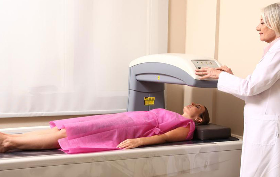
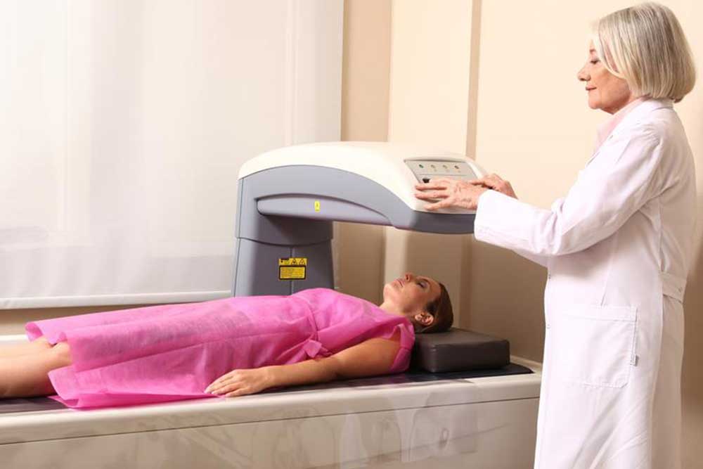
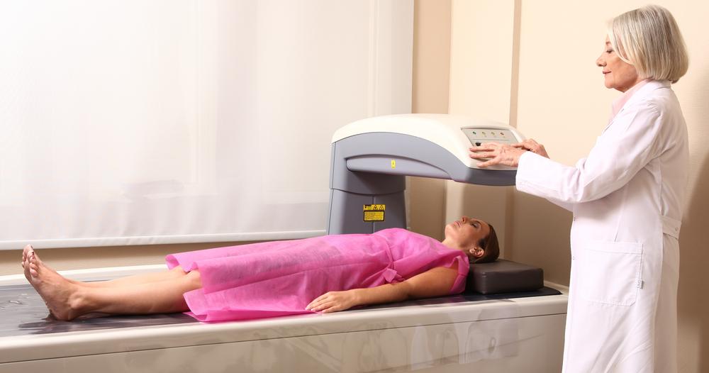
Monitoring BMD through repeat testing offers valuable clinical insights, provided tests are accurately performed and interpreted by knowledgeable clinicians. The most reliable method for diagnosing and tracking osteoporosis is dual-energy X-ray absorptiometry (DXA) scans of the spine, hip, or forearm. DXA has demonstrated a strong link between bone density and bone strength, with excellent accuracy and minimal radiation exposure.
DXA results are often expressed as a T-score, comparing a patient's BMD with that of a healthy young adult. Based on WHO criteria, postmenopausal women’s BMD is classified as normal, osteopenia, or osteoporosis. Developing standardized methods help determine cost-effective treatment thresholds based on T-scores combined with other fracture risk factors.