Understanding Bone Density Testing and Its Importance
This article provides a comprehensive overview of bone density testing, its significance in diagnosing and managing osteoporosis, and the factors influencing bone health. It explains how the scan works, who needs it, and its role in treatment monitoring, emphasizing the importance of early detection for preventing fractures and maintaining bone strength.
Sponsored
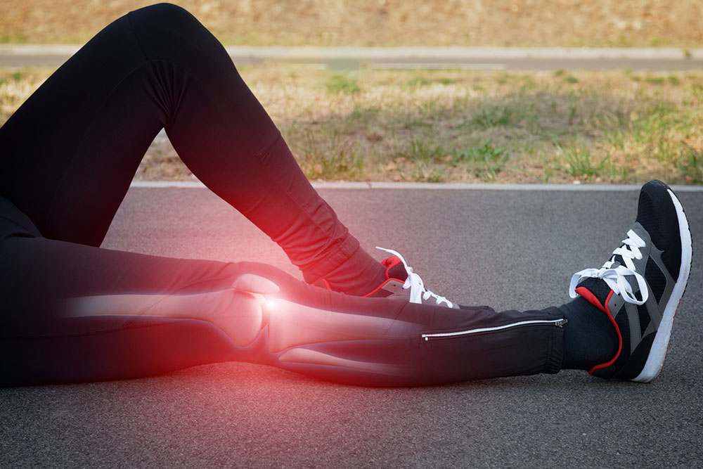
Osteoporosis is a condition characterized by weakened bones, increasing the risk of fractures in areas such as the spine, wrist, and hips. Though more common in women, men can also develop this disease. In the U.S., approximately 10 million people are affected—8 million women and 2 million men—and an additional 34 million are at high risk due to decreased bone mass, known as osteopenia.
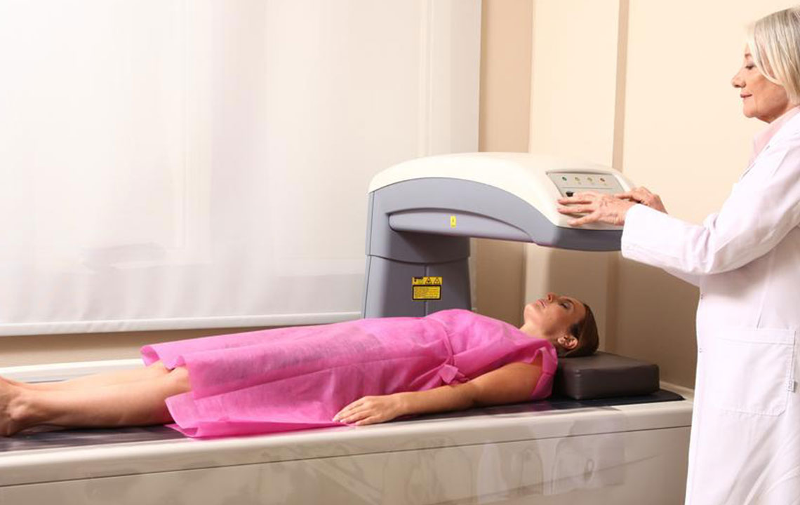
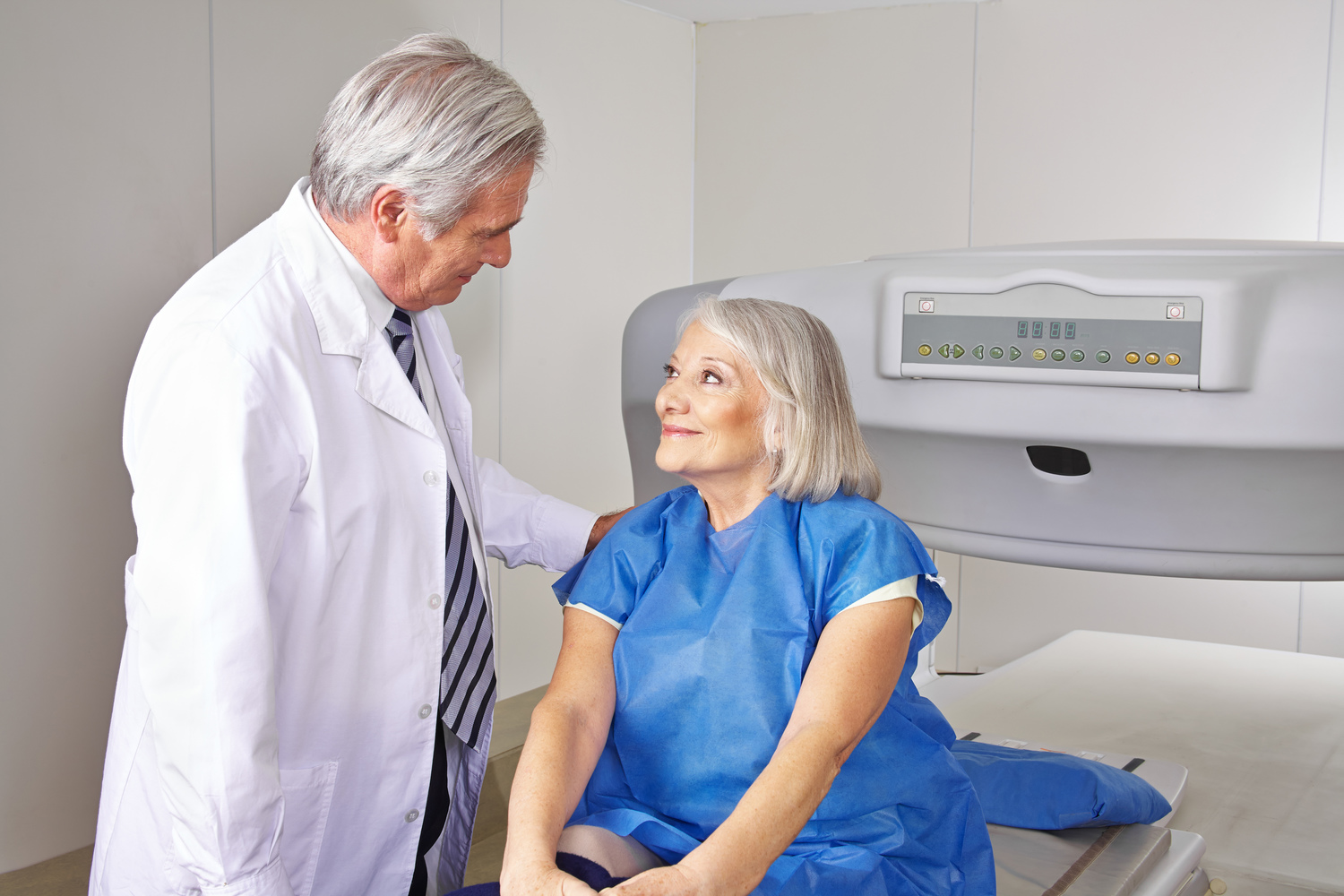
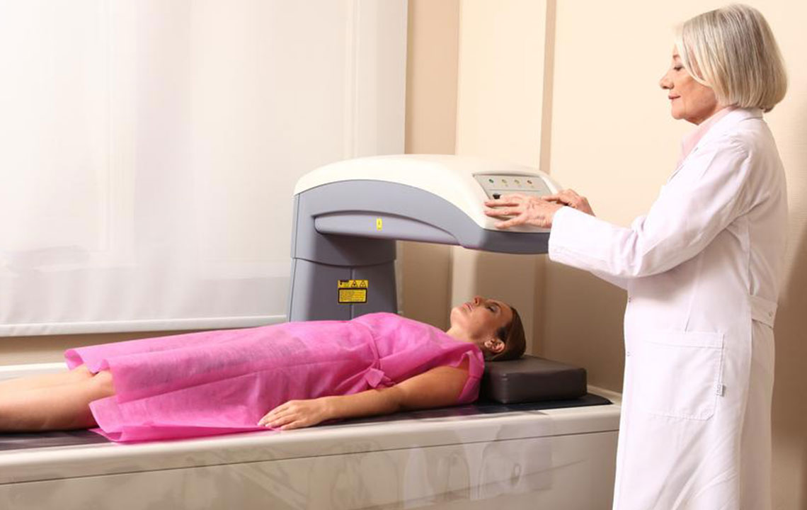
A bone density scan is a quick, non-invasive method used to diagnose osteoporosis and monitor treatment progress. Medical professionals may recommend this test based on specific risk factors.
Height reduction: Losing more than 1.6 inches in height can signal spinal fractures caused by osteoporosis.
Bone fractures: Fragile bones are more susceptible to breaking.
Medications: Long-term use of certain drugs can impair bone formation, leading to osteoporosis.
Organ transplants: Transplant procedures can compromise bone health by disrupting normal rebuilding processes.
Hormonal changes: Post-menopause, women experience a natural decline in estrogen, increasing osteoporosis risk. Similarly, treatments for cancers like prostate and breast can reduce hormone levels, affecting bone strength in both men and women.
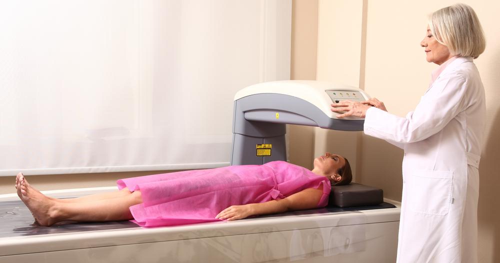
The test involves a low-dose X-ray to detect decreased bone mineral content, focusing on the hip and spine. When the hip isn’t accessible, forearm scans are used, especially for patients with conditions like hyperthyroidism. For younger individuals under 60, clinicians typically recommend hip scans. The procedure takes about 15 minutes, with patients lying flat on a table for 5-8 minutes while detailed 3D images are generated. These images help assess bone health, aging effects, and other bone-related conditions.