Understanding Bone Density Testing: A Guide
Learn about bone density testing, a vital screening method for osteoporosis risk. This guide explains when and why doctors recommend these quick, non-invasive scans to detect and monitor bone health. Discover how the procedure works, the conditions prompted for testing, and the significance of early detection in preventing fractures and bone loss. Appropriate for patients and caregivers, this overview highlights the importance of maintaining strong bones through timely diagnosis and management.
Sponsored
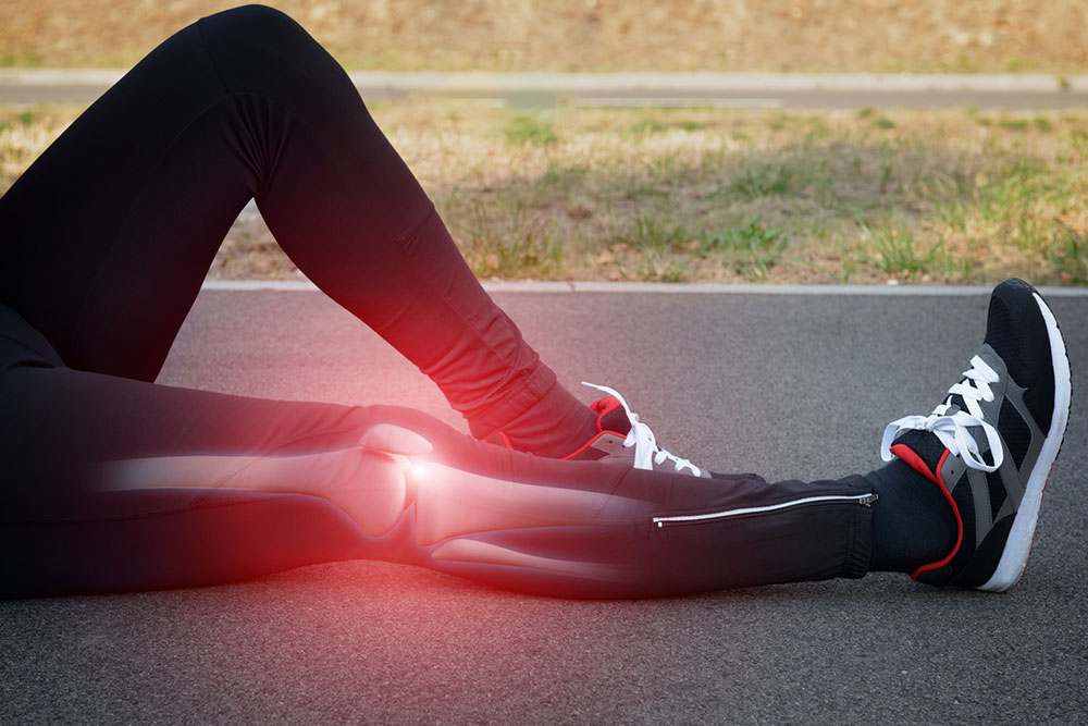
Osteoporosis is a condition marked by weakened bones, leading to an increased risk of fractures, especially in the spine, wrist, and hip. It predominantly affects women, but men are also susceptible. In the United States, around 10 million individuals are affected, including 8 million women and 2 million men, with an additional 34 million at high risk due to low bone mass, known as osteopenia.
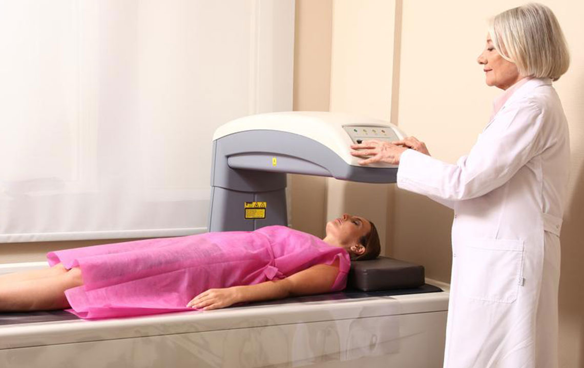
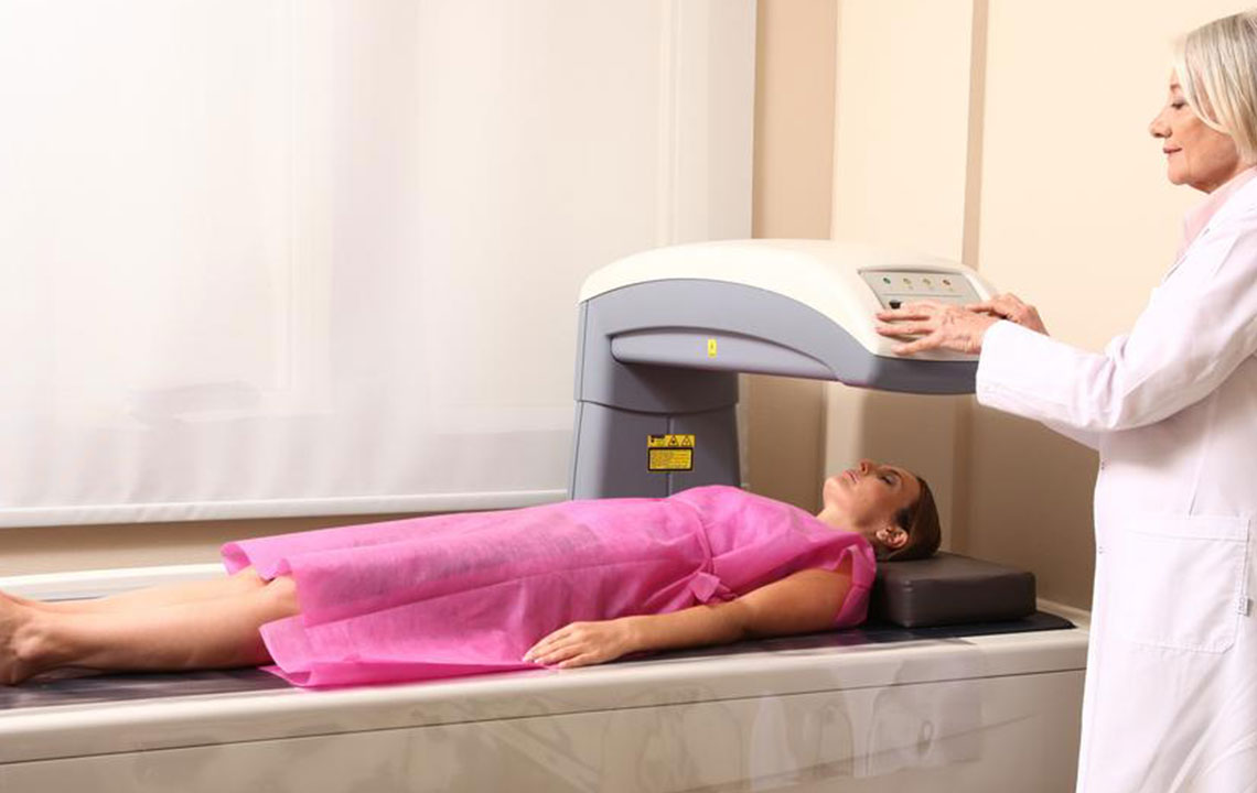
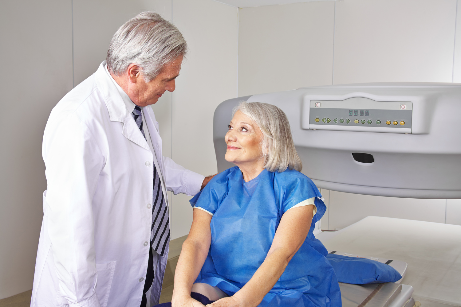
A bone density examination is a quick, non-invasive method used to detect osteoporosis risk and track treatment progress.
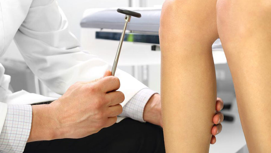
Doctors may recommend a bone density test if you experience:
Height reduction: Losing at least 1.6 inches may signal vertebral fractures caused by osteoporosis.
Bone fractures: Fragile bones are more prone to breaking.
Long-term steroid use: Extended use of medications like prednisone can impair bone renewal, increasing osteoporosis risk.
Organ transplants: Transplant recipients often face higher osteoporosis risk due to immunosuppressant medications affecting bone health.
Hormonal changes: Postmenopausal women experience a natural decline in estrogen, affecting bone strength. Similarly, treatments for prostate or other cancers can lower testosterone, raising osteoporosis risk in men.
A low-dose X-ray scan can detect signs of decreasing bone density and mineral loss. The scan focuses on the hip and spine, with alternative options like forearm scans for those with hyperthyroidism or inability to scan the hip. For those under 60, only hip scans are typically recommended.
Bone density is measured using central or peripheral devices. Central scanners assess the spine, hip, and total body, while peripheral scanners evaluate areas like the heel, shin, wrist, or finger. The procedure lasts around 15 minutes, with the patient lying on a table for 5-8 minutes. The resulting detailed 3D images provide insights into bone health and age-related changes beyond osteoporosis.