Understanding Osteoarthritis: Causes and Joint Changes
Osteoarthritis is a prevalent joint disease causing cartilage breakdown, bone changes, and inflammation. It mainly affects knees, hips, and hands, especially in women and obese individuals. The condition involves complex pathological changes in cartilage, subchondral bone, and synovial membranes, leading to joint pain, stiffness, and reduced mobility. Understanding these mechanisms helps in early diagnosis and targeted treatment to manage symptoms and slow progression.
Sponsored

Osteoarthritis (OA) is the leading form of arthritis worldwide, characterized by the gradual deterioration of joint cartilage and bone beneath it. Affecting nearly 15% of the global population, OA most commonly impacts the knees, hips, and smaller joints like the hands. The risk of developing OA during one’s lifetime ranges from 40% to 47%, with higher prevalence in obese individuals and athletes. OA results from a complex interplay of genetic, metabolic, biomechanical, and biochemical factors.
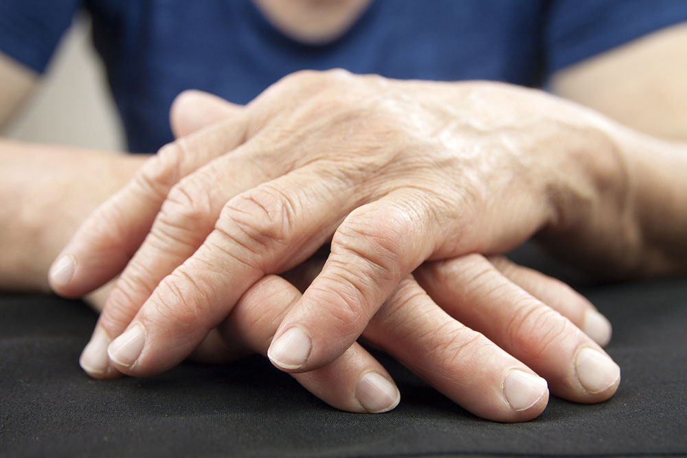
The disease can affect virtually any joint, with 7% to 19% of adults experiencing it in their hands or knees. Women are more frequently affected by OA.
OA involves three key joint components: cartilage, subchondral bone, and synovial tissue.
Cartilage Damage
The articular cartilage covers the ends of bones that form the joint. It relies on an intricate matrix of proteins and molecules, produced by specialized cells called chondrocytes, which maintain the cartilage's integrity. An imbalance between cartilage breakdown and repair leads to OA. This imbalance may result from trauma-induced inflammation, activating enzymes that produce wear particles. These particles provoke immune responses, release inflammatory cytokines, and decrease cartilage matrix components like type II collagen. Over time, cartilage becomes weak, soft, and thin, resulting in fissures and exposure of the subchondral bone.
Subchondral Bone Changes
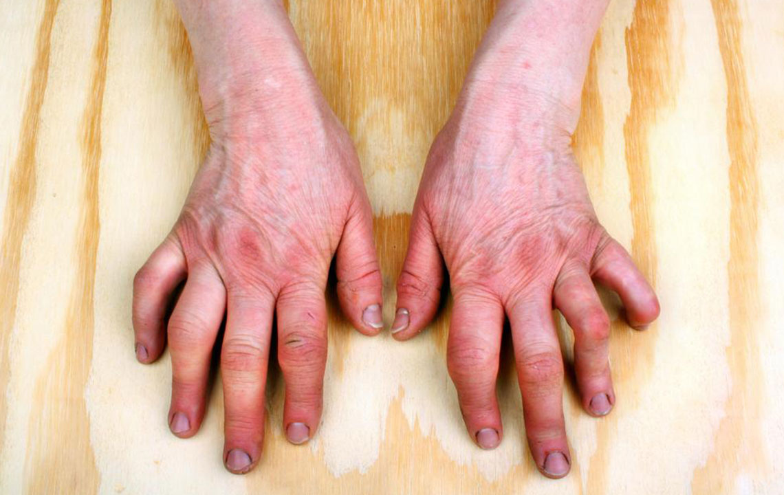
The subchondral bone beneath the cartilage absorbs shock during movement. During OA, remodeling processes lead to bone marrow lesions, cyst formation, and osteophyte development—bony projections at joint edges. Advanced OA causes significant bone loss, necrosis, and abnormal mineralization, weakening the joint’s ability to withstand mechanical forces and contributing to joint deformation.
Synovial Membrane and Meniscus Alterations
The synovial membrane lines the joint’s interior, producing fluid that lubricates joint movement. In OA, it thickens and becomes inflamed, releasing cytokines like interleukins and TNFα, which contribute to pain and further joint damage. The meniscus, a cartilage structure stabilizing the joint, often degenerates or tears in OA, worsening joint instability and degeneration.