Understanding Bone Mineral Density Testing: Methods and Significance
Bone mineral density testing is vital for early osteoporosis detection, utilizing DXA or QCT methods to assess bone health. The procedure is safe, quick, and informative, helping prevent fractures and improving treatment strategies.
Sponsored

Assessing bone strength is crucial for diagnosing conditions like osteoporosis. Bone mineral density (BMD) testing provides a measure of bone quality, typically focusing on the spine or hips. Before these tests, osteoporosis was often diagnosed only after fractures occurred. Now, early detection allows for better preventive care.
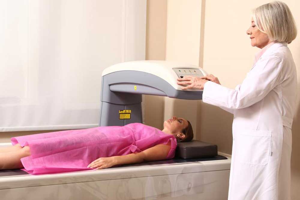
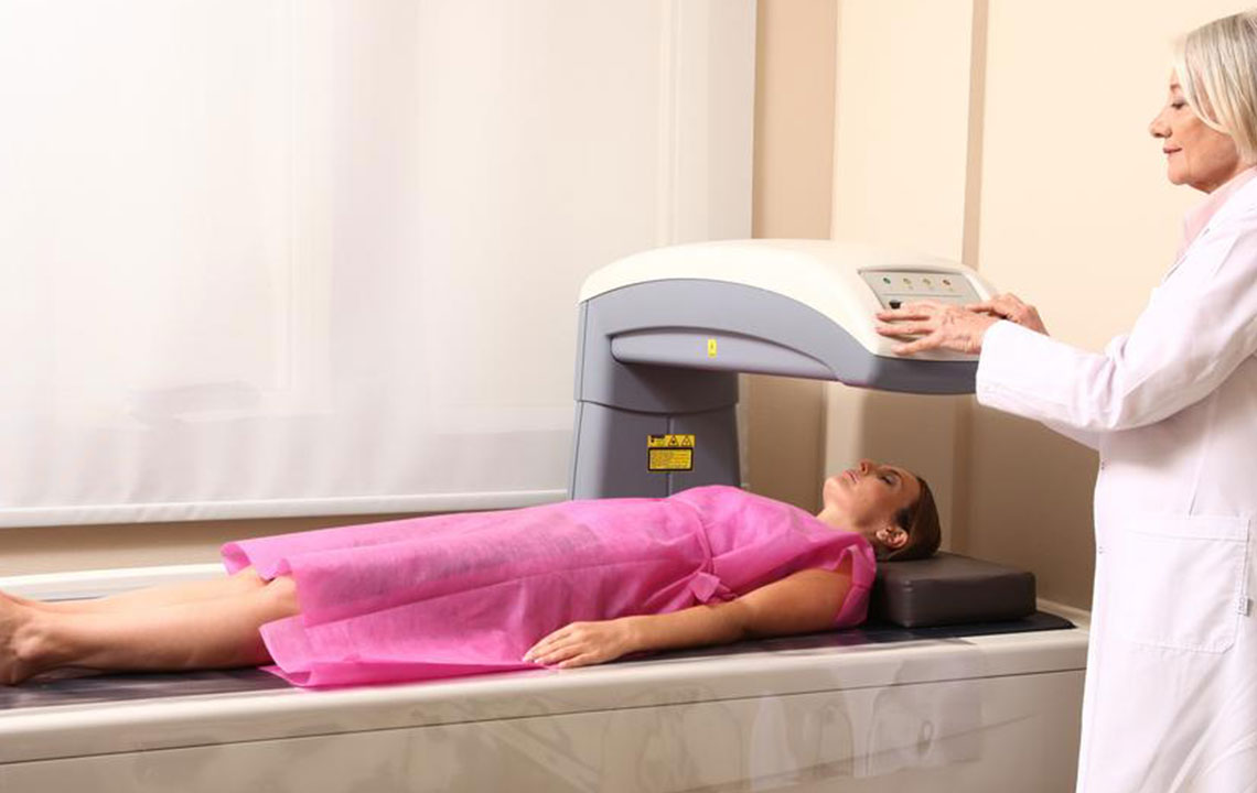
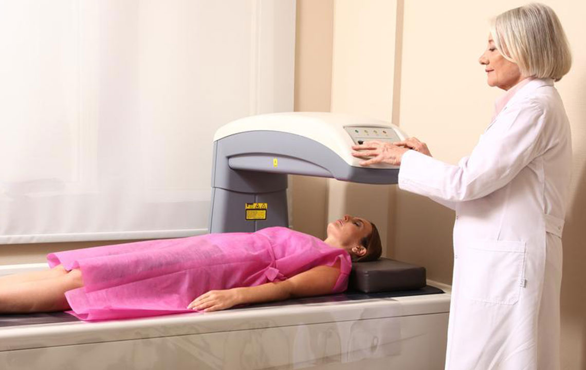
The process resembles an X-ray, where a person lies on a platform while a small arm measures radiation exposure. This safe procedure takes around 10 to 30 minutes and targets high-risk bones. Peripheral devices exist but are less reliable for diagnosis.
Methods of BMD Testing
DXA Scan uses minimal radiation to image bones, calculating loss from these images. No special prep is needed—just avoid jewelry and wear loose clothing. Refrain from calcium supplements 24 hours prior.
QCT Scan employs a standard CT scanner to analyze bone density, especially useful for patients with scoliosis. It provides faster results and assesses soft bone tissue around the spine, often affected early in osteoporosis.
Results are expressed as T-score and Z-score.
T-score compares your bone density with what is typical for your sex: -1 or above indicates normal bone health; between -1 and -2.5 suggests low bone density; -2.5 or below confirms osteoporosis.
Z-score reflects how your bone density compares to peers of the same age and sex. A Z-score lower than -2 indicates another factor beyond aging may be causing bone loss.