Understanding Catheter-Directed Thrombolysis for Venous Clot Removal
Discover the essentials of catheter-directed thrombolysis, a minimally invasive technique for dissolving blood clots in veins. Learn about the procedure, preparation, equipment, benefits, and risks. A safe alternative to surgery, it restores blood flow efficiently, reducing recovery time and complications associated with deep vein thrombosis.
Sponsored

Overview of Catheter-Directed Thrombolysis as a Treatment for Venous Clots
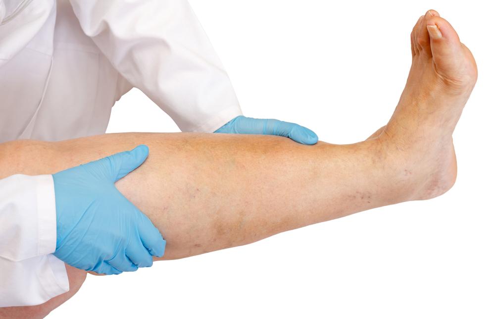
Deep vein thrombosis (DVT) occurs when a blood clot forms deep within a vein, often in the legs or thighs. Symptoms include skin redness, swelling, and tenderness over affected veins. It is more common in individuals over 50 or those with conditions affecting blood flow. Recognizing DVT early is crucial to prevent complications such as pulmonary embolism.
What is catheter-directed thrombolysis?
This minimally invasive procedure uses a catheter guided by X-ray imaging to dissolve blood clots within veins.
It restores normal blood flow, preventing tissue or organ damage caused by reduced circulation.
Advantages of this method
It is a safe, effective option for treating venous clots.
No surgical incision is necessary.
It helps improve blood circulation efficiently.
The procedure avoids invasive surgery, reducing recovery time.
Patients typically experience shorter hospital stays compared to open procedures.
Preparation Steps for the Procedure
Inform your doctor about any current or past medications.
Disclose existing health conditions or medical issues.
Pregnant women should inform their healthcare provider about pregnancy status.
Pre-procedure blood tests assess kidney function and clot status.
Follow any additional instructions provided by your doctor, including medication adjustments.
Procedure Process
The targeted vessel is identified using contrast dye and X-ray guidance.
A thin catheter is inserted through a small skin opening and navigated to the clot's location.
The blood clot is then dissolved either through direct medication delivery or mechanical fragmentation.
Equipment Used
A slender plastic catheter approximately spaghetti-thin.
X-ray imaging systems, medications or mechanical devices for clot dissolution.
Additional tools include monitors, ultrasound machines, and IV lines, depending on the treatment plan.
Understanding Treatment Results
The radiologist interprets post-procedure outcomes to determine success.
Follow-up treatments may be needed if tissue damage occurs.
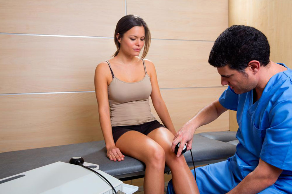
Carried out through physical examinations, blood tests, or imaging studies.
Potential Risks
Infection risk is low, approximately 0.1%.
Possible allergic reactions, though rare.
Potential vessel injury, with a very low incidence.
Bleeding at different sites may occur.
Patients with kidney issues could experience rare kidney damage.
Blood clots may dislodge and require additional intervention.